

Selecting the Best Neuronavigation and Advanced Brain Surgery Centres in India for Nigerian Patients
Neuronavigation is often described as GPS for brain surgery. Unlike GPS, its map stops updating the moment the terrain actually starts to move.
Neuronavigation registers a patient's preoperative brain scan to their actual anatomy, allowing a surgeon to see, in real time, exactly where their instruments sit relative to a tumour and critical surrounding structures. What is genuinely underexplained to most patients is that this map is built before surgery begins, and the brain itself physically moves once the skull is opened, a well- documented phenomenon called brain shift. Understanding this limitation, and what a specific centre does to correct for it, matters more than the general reassurance that "advanced neuronavigation" is being used. This guide sets out that specific, measurable limitation, the two main technologies used to correct for it, and why this particular evaluation carries a meaningfully different balance between surgeon and hospital than several other guides in this series.
Should you even be reading this guide? If complex brain tumour surgery using neuronavigation has genuinely been recommended and confirmed as appropriate, this guide will help you evaluate the specific centre's technology and team. This series' main Neuronavigation guide covers the broader treatment landscape.
Healing Journeys of Nigerian Patients


Key Takeaways
- The guide's central message is that neuronavigation is valuable but not automatically accurate throughout brain surgery because the brain can move after the skull is opened.
- This movement, known as brain shift, can result from cerebrospinal fluid drainage, gravity, swelling and tumour removal. The page 2 illustration notes displacement exceeding 3 mm at the deep tumour margin in 66% of documented cases.
- The guide identifies two main technologies for correcting the navigation map during surgery: intraoperative MRI and intraoperative ultrasound.
- The page 3 comparison describes intraoperative MRI as offering gold-standard accuracy, while ultrasound provides real-time, repeatable imaging at lower resolution.
- The guide explains that neither technology is universally superior. The important question is what technology the centre actually has and how routinely it is used.
- Brain-shift compensation is particularly important for deep-seated tumours, tumours near critical structures and lengthy operations, while its impact may be smaller for small, superficial and easily accessible tumours.
- The page 4 evaluation gives 53% weight to the hospital's brain-shift compensation technology and 47% to the surgeon's judgement and awareness of neuronavigation limitations.
- Nigerian patients should ask specifically which compensation technology will be used during their surgery, rather than accepting a general statement that the centre offers “advanced neuronavigation.”
- The guide also recommends asking about the surgeon's specific experience with tumours in the patient's anatomical location, rather than relying only on overall neurosurgical experience.
- Four warning signs include claiming neuronavigation remains fully precise throughout surgery, inability to identify brain-shift compensation technology, no discussion of brain shift for the specific tumour and vague answers about routine equipment use.
Quick Facts
- Treatment
- Neuronavigation-Assisted Brain Surgery
- Country
- India
- Patients
- Patients from Nigeria
- Key Specialist
- Neurosurgeon / Advanced Brain Surgery Specialist
- Main Technology
- Neuronavigation With Brain-Shift Compensation
- Primary Limitation
- Brain Shift After Skull Opening
- Deep Margin Finding
- Displacement Exceeding 3 mm in 66% of Documented Cases
- Compensation Options
- Intraoperative MRI or Intraoperative Ultrasound
- Intraoperative MRI
- Higher Accuracy / Gold-Standard Imaging
- Intraoperative Ultrasound
- Real-Time and Repeatable Imaging
- Technology Evaluation
- 53% Weight
- Surgeon Evaluation
- 47% Weight
- Important Check
- Confirm Specific Compensation Technology
- Usage Check
- Ask How Often the Technology Is Actually Used
- Surgeon Check
- Confirm Experience With the Specific Tumour Location
- Pre-Travel Records
- Full Imaging and Confirmed Diagnosis
- Important Warning
- Do Not Assume Neuronavigation Remains Fully Accurate Throughout Surgery
- Follow-Up
- Nigerian Neurologist or Oncologist With Appropriate Imaging
- Author
- Dr. Dheeraj Bojwani
- Experience
- 24+ Years
In Brief
For Nigerian patients considering neuronavigation-assisted brain surgery in India, the guide recommends evaluating how the centre manages brain shift rather than simply confirming that neuronavigation is available. Patients should ask whether intraoperative MRI or ultrasound will be used, how routinely the technology is employed, how brain shift affects their specific tumour location and what experience the surgeon has with that type of tumour. Before travelling, patients should complete their diagnosis and imaging, confirm the centre's technology and establish a coordinated follow-up plan with a Nigerian neurologist or oncologist.
START HERE
The map stops updating the moment the skull opens
Understanding this specific, measurable limitation is worth doing before evaluating any centre's neuronavigation claims.
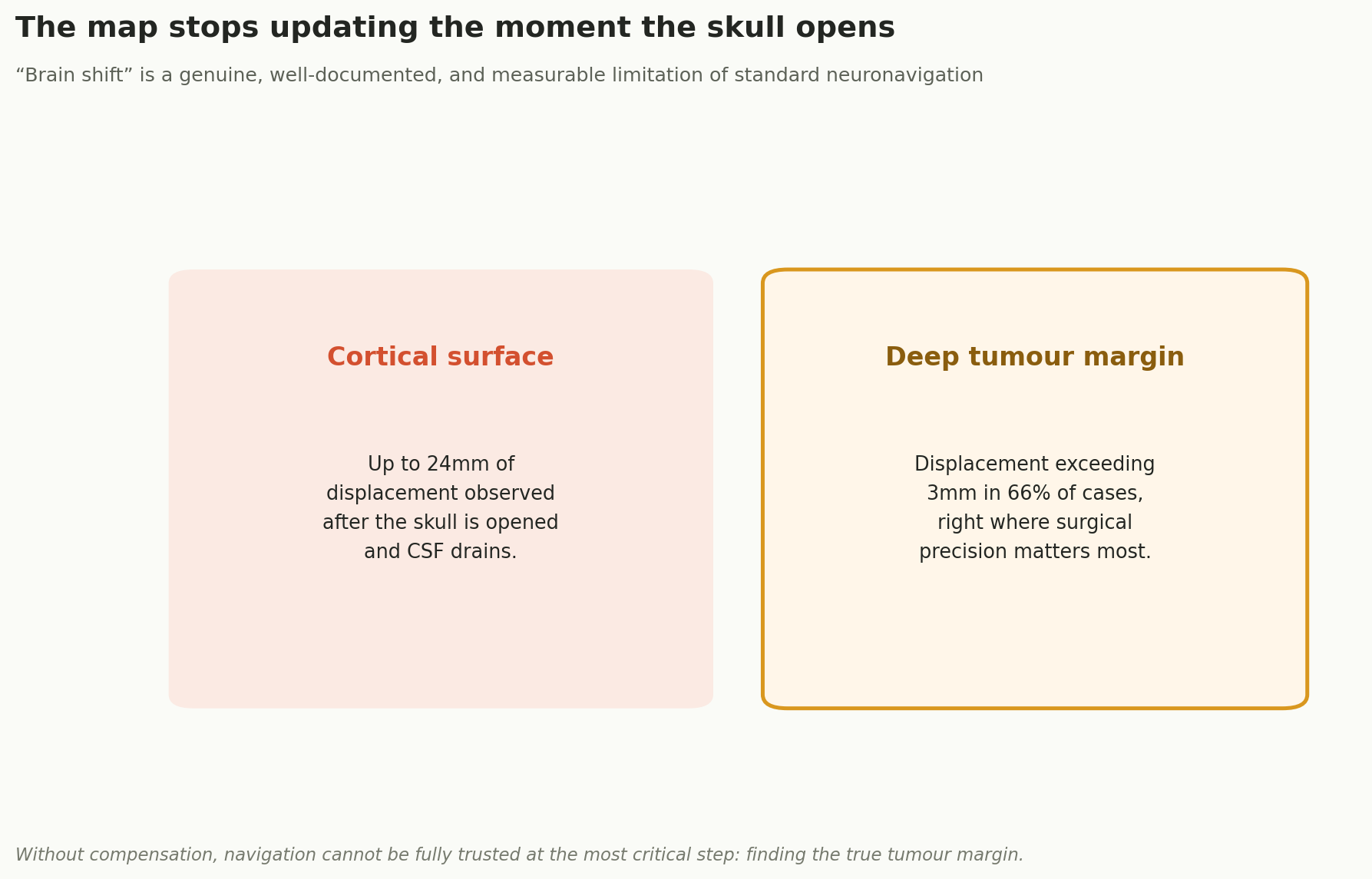
Without compensation, navigation cannot be fully trusted at the most critical step: finding the true tumour margin.
Brain shift is caused by cerebrospinal fluid drainage, gravity, swelling, and the physical removal of tumour tissue during surgery, and it is not a rare or minor effect; published research has measured displacement at the deep tumour margin, precisely where surgical precision matters most, in the majority of cases studied. This does not mean neuronavigation is unreliable throughout an operation; it means its accuracy genuinely depends on whether, and how, this shift is actively corrected for as surgery proceeds.
It is worth being specific about why this matters practically, not just technically. Without correction, a surgeon relying on the original preoperative map late in a procedure may believe they are still working precisely at the tumour margin, when the actual anatomy has already shifted meaningfully away from that plan. This is precisely the scenario published research warns cannot be fully trusted without active compensation.
How This Gets Corrected
Two genuinely different ways to correct the map mid-surgery
Understanding these options helps frame a specific, useful question for any prospective centre.
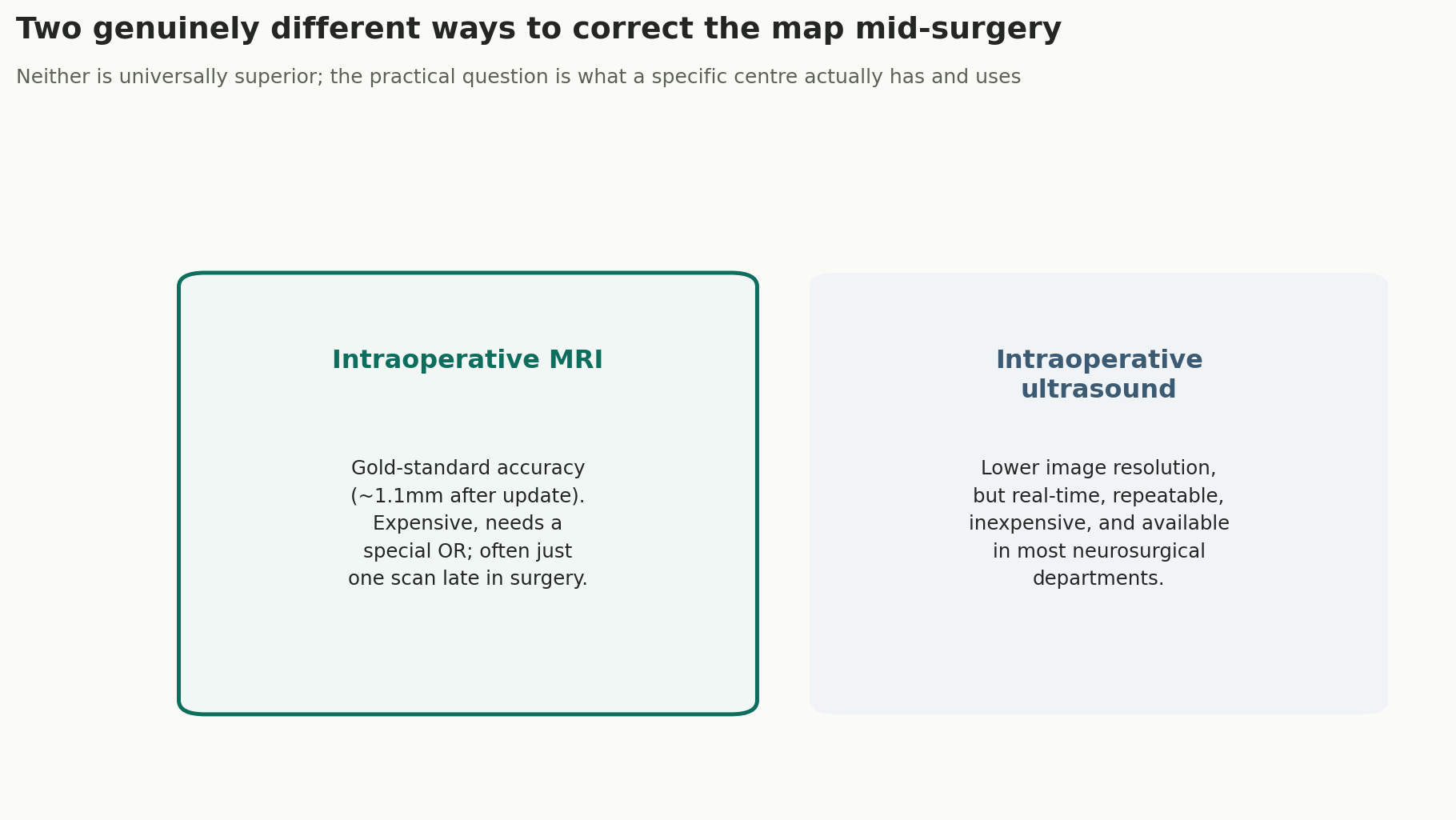
Neither is universally superior; the practical question is what a specific centre actually has and uses.
A centre without either technology is not necessarily unsafe for straightforward cases, but for complex tumours, particularly those near critical structures, the absence of any brain-shift compensation method is a genuinely relevant limitation worth understanding before committing to a specific programme.
Cost and infrastructure requirements explain much of why these two technologies remain unevenly distributed even among otherwise well-regarded neurosurgical centres. A hospital's decision to invest in one, both, or neither reflects genuine resource constraints as much as clinical philosophy, which is exactly why this is a question worth asking directly rather than assuming any advanced-sounding facility has already solved it.
A note on why this matters more for some tumours than others. Brain shift's practical impact depends heavily on tumour location and surgical duration. A small, superficial, easily accessible tumour is less affected than a deep-seated tumour near critical structures in a lengthy operation. Asking a surgeon directly how brain shift specifically affects your tumour's location is a genuinely useful, case-specific question.
THE BALANCE
Trust the surgeon who trusts the technology less blindly
Given how much correcting for brain shift depends on specific equipment a hospital either has or does not have, this evaluation weights slightly toward the centre.
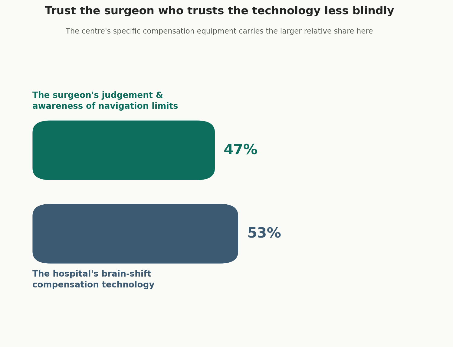
The centre's specific compensation equipment carries the larger relative share here.
The surgeon's own judgement remains genuinely essential, particularly their willingness to acknowledge navigation's limitations honestly rather than treating it as infallible. But whether brain-shift compensation is available at all is fundamentally an equipment question, not a skill question, which is why the centre's specific technology carries the slightly larger relative share of this particular evaluation. This is a genuinely different pattern than several more surgeon-dominated procedures elsewhere in this series, reflecting how much this specific limitation is constrained by available infrastructure rather than individual skill alone.
THE VETTING CONVERSATION
What to actually ask
Questions for the surgeon
- How does brain shift specifically affect my tumour's location? A thoughtful, specific answer engages honestly with the limitation.
- What compensation technology will be used during my specific surgery? This should be a clear, confident answer.
- What is your specific experience operating on tumours in this location? Ask specifically, not just general neurosurgical volume.
- How do you decide when navigation data needs updating during surgery? This should reflect genuine, active awareness, not passive trust in the system.
Questions for the hospital
- Does this centre have intraoperative MRI, intraoperative ultrasound, or neither? This should be a specific, verifiable answer.
- How often is compensation technology actually used during surgery, not just available? Availability and actual use are different facts.
- What is the centre's own outcome data for tumours in this specific location? Specific, tracked figures matter more than general reputation.
- What is the plan for follow-up imaging and care once I return to Nigeria? This should be coordinated before you travel.
Nigeria-specific Considerations
Why this question matters even more from a distance
With advanced neurosurgical infrastructure, including intraoperative MRI, remaining genuinely limited across much of Sub-Saharan Africa, most Nigerian patients evaluating this treatment are doing so from a distance, often trusting a centre's own description of its capabilities. This makes directly confirming specific brain-shift compensation technology, rather than accepting general reassurance about "advanced neuronavigation," a particularly important verification step for Nigerian patients specifically.
Four warning signs worth taking seriously
- A centre describing neuronavigation as fully precise throughout surgery. This overstates what the technology genuinely does without compensation.
- Inability to confirm specific brain-shift compensation technology. This should be a clear, specific answer.
- No discussion of how brain shift affects your particular tumour location. This should be raised proactively for complex cases.
- Vague answers about actual, routine use of available equipment. Availability alone does not confirm genuine, consistent use.
A practical order of operations
- Get a confirmed diagnosis and full imaging to understand your tumour's specific location.
- Ask directly how brain shift is expected to affect your particular case.
- Confirm which specific compensation technology a centre has, and how routinely it is used.
- Ask the surgeon-side and hospital-side questions above before committing to a programme.
- Travel, surgery, and a follow-up imaging schedule appropriate to your specific case.
- Establish a coordinated follow-up relationship with a Nigerian neurologist or oncologist.
Straight Answers for Nigerian Patients about Neuronavigation and Advanced Brain Tumor Surgery in India
Does neuronavigation stay accurate throughout surgery?
Not automatically. Brain shift progressively degrades its accuracy once the skull is opened, with deep tumour margin displacement exceeding 3mm in 66 percent of documented cases.
How is brain shift corrected?
Intraoperative MRI offers gold-standard accuracy but is expensive and often limited to one scan. Intraoperative ultrasound offers real-time, repeatable, more widely available imaging at lower resolution.
Is intraoperative MRI always better than ultrasound?
Not necessarily. MRI offers higher accuracy but limited, infrequent use; ultrasound offers real-time, repeatable updates throughout surgery at considerably lower cost.
Why does the hospital's technology matter more than the surgeon here?
Correcting for brain shift depends on specific equipment a hospital either has or does not have, a factor less within an individual surgeon's control than in many other procedures.
What is a realistic red flag when choosing a centre?
Describing neuronavigation as fully precise without acknowledging brain shift, or inability to confirm specific compensation technology, are genuine warning signs.
A closing word
Neuronavigation is a genuine, valuable surgical tool, and its limitations do not diminish its value; they simply mean it should be understood accurately rather than marketed as infallible. A centre honest about brain shift, and equipped to correct for it, is offering something meaningfully different from one that presents "advanced neuronavigation" as a solved problem. In twenty-four years of this work, the families who navigate this best ask specifically about brain-shift compensation technology, understand how it applies to their particular tumour's location, and treat a surgeon's honest acknowledgement of navigation's limits as a genuine marker of expertise, not a source of doubt.
Sources
- 🌐 Neuronavigation in glioma resection: current applications, challenges, and clinical outcomes. Frontiers in Surgery, 2024
- 🌐 Use of virtual magnetic resonance imaging to compensate for brain shift during image-guided surgery. PMC
- 🌐 Quantification of, visualization of, and compensation for brain shift using intraoperative magnetic resonance imaging. PubMed
- 🌐 What neurosurgeons need to see: synthetic intra-operative MRI from ultrasound for brain-shift compensation, 2026
- 🌐 High Commission of India, Abuja — Medical and Medical Attendant Visa requirements
Frequently Asked Questions by Nigerians about Neuronavigation and Advanced Brain Tumor Surgery in India
Does neuronavigation remain accurate throughout brain surgery?
Not automatically. The guide explains that brain shift occurs after the skull is opened, progressively reducing the accuracy of the original preoperative navigation map.
What is brain shift?
Brain shift is movement of brain tissue caused by factors such as cerebrospinal fluid drainage, gravity, swelling and removal of tumour tissue during surgery.
How significant can brain shift be?
The page 2 illustration reports deep tumour-margin displacement exceeding 3 mm in 66% of documented cases, showing why compensation can matter during complex surgery.
How is brain shift corrected during surgery?
The guide identifies intraoperative MRI and intraoperative ultrasound as two different approaches for updating the surgical map during the operation.
Is intraoperative MRI always better than intraoperative ultrasound?
Not necessarily. MRI provides higher accuracy, while ultrasound provides real-time, repeatable updates and is generally more accessible and less costly.
Which brain tumours may require greater attention to brain-shift compensation?
The guide explains that the impact is greater for deep-seated tumours, tumours close to critical structures and lengthy operations than for small, superficial tumours.
What should Nigerian patients ask a neurosurgeon about neuronavigation?
They should ask how brain shift affects their specific tumour location, which compensation technology will be used and what experience the surgeon has with tumours in that location.
What should Nigerian patients ask the hospital about its technology?
They should confirm whether the centre has intraoperative MRI, intraoperative ultrasound or neither, and how frequently the available technology is actually used during surgery.
What are the major warning signs when choosing a neuronavigation centre?
Warning signs include claiming that neuronavigation is fully precise throughout surgery, inability to identify compensation technology, no discussion of brain shift and vague information about actual equipment use.
What should Nigerian patients arrange before returning home after brain surgery?
They should establish a coordinated follow-up relationship with a Nigerian neurologist or oncologist and confirm the appropriate follow-up imaging schedule for their specific case.
Page Summary
This guide explains how Nigerian patients can select neuronavigation and advanced brain surgery centres in India, focusing on the important limitation of brain shift during surgery. The page 2 illustration shows that brain movement can affect the accuracy of the original navigation map, with deep tumour-margin displacement exceeding 3 mm in 66% of documented cases. The page 3 comparison presents intraoperative MRI and intraoperative ultrasound as the two main approaches for updating or compensating for the navigation map, while noting that each has different advantages. The page 4 evaluation gives 53% weight to the hospital's brain-shift compensation technology and 47% to the surgeon's judgement and awareness of navigation limitations. The guide also covers tumour-specific assessment, actual equipment use, Nigerian follow-up and warning signs when evaluating a centre.
Citation Block
| Topic | Information |
|---|---|
| Topic Information | Selecting the Best Neuronavigation and Advanced Brain Surgery Centres in India for Nigerian Patients for Patients from Nigeria |
| Treatment | Neuronavigation-Assisted Brain Surgery |
| Intended Audience | Patients from Nigeria |
| Key Specialist | Neurosurgeon / Advanced Brain Surgery Specialist |
| Primary Technology | Neuronavigation With Brain-Shift Compensation |
| Main Limitation | Brain Shift After the Skull Is Opened |
| Deep Tumour Margin Finding | Displacement Exceeding 3 mm in 66% of Documented Cases |
| Compensation Options | Intraoperative MRI and Intraoperative Ultrasound |
| Intraoperative MRI | Gold-Standard Accuracy; Expensive and Less Frequently Available |
| Intraoperative Ultrasound | Lower Resolution but Real-Time and Repeatable |
| Evaluation Weighting | 53% Hospital Technology; 47% Surgeon Judgement |
| Technology Check | Confirm Which Brain-Shift Compensation Technology Is Available |
| Usage Check | Confirm How Routinely the Technology Is Used During Surgery |
| Surgeon Check | Ask About Experience With Tumours in the Specific Location |
| Case Assessment | Consider Tumour Location and Expected Surgical Duration |
| Important Warning | Do Not Treat Neuronavigation as Fully Precise Throughout Surgery |
| Nigeria-Specific Check | Independently Verify Technology Rather Than Relying on General “Advanced” Claims |
| Pre-Travel Requirement | Confirm Diagnosis and Complete Brain Imaging |
| Patient Journey | Diagnosis/imaging → brain-shift assessment → technology verification → surgeon/centre evaluation → travel/surgery → follow-up imaging |
| Follow-Up | Coordinated Care With a Nigerian Neurologist or Oncologist |


Our Mission Statement
Our mission is to place patients at the centre of every healthcare decision by providing trustworthy, evidence-based, and unbiased medical information. We believe that informed patients make better decisions. Backed by personalised guidance from our experienced advisory team, we help patients understand their treatment options, compare them objectively, and confidently choose the path that best suits their medical needs and personal circumstances.
Patient Testimonials from Nigeria

Ready to Take the First Step?
Share your Medical Reports with our Healthcare Managers Today and Get a FREE CONSULTATION, a Personalized Treatment Plan, and Complete Support from Arrival to Recovery.
Get Your Free ConsultationAreas We Serve
This resource has been thoughtfully prepared for patients from Nigeria who are considering medical treatment in India. We also welcome patients from the wider region in the countries mentioned below-
We assist patients from:
- Benin
- Niger
- Chad
- Cameroon
- Algeria
- Egypt
- Libya
- Morocco
- Sudan
- Tunisia
- Gambia
- Ghana
- Burkina Faso
- Cabo Verde
- Côte d'Ivoire
- Equatorial Guinea
- São Tomé and Príncipe
Many of the insights, treatment pathways, hospital recommendations, travel guidance, and patient support services described here are equally relevant and may be used as a reference when planning treatment in India.
From Nigeria to India: Your Complete Patient Support Guide
- Artificial Disc Replacement in India for Nigerian Patients
- Weight Loss and Bariatric Surgery in India for Nigerian Patients
- Bone Marrow Transplant in India for Nigerian Patients
- Brain Tumour Surgery in India for Nigerian Patients
- Breast Cancer Treatment and Surgery in India for Nigerian Patients
- Cancer Treatment and Surgery in India for Nigerian Patients
- Cardiac Arrhythmia and Heart Pacemaker Treatment in India for Nigerian Patients
- Cardiac Surgery in India for Nigerian Patients
- Colorectal Cancer Treatment and Surgery in India for Nigerian Patients
- Complex Orthopaedic and Joint Revision Surgery in India for Nigerian Patients
- Cornea Transplant and Advanced Eye Surgery in India for Nigerian Patients
- CyberKnife Treatment in India for Nigerian Patients
- Gamma Knife Treatment in India for Nigerian Patients
- Heart Failure Treatment and Surgery in India for Nigerian Patients
- Hip Surgery in India for Nigerian Patients
- HIPEC Cancer Surgery in India for Nigerian Patients
- IVF and Fertility Treatment in India for Nigerian Patients
- Joint Arthroscopy and Sports Medicine in India for Nigerian Patients